ANALYTICAL OFFER
ADVANCED GEOCHRONOLOGY
U-Pb dating of zircons and others U-Pb-Th minerals such as zircon, monazite, titanite, perovskite, allanite, rutile, baddeleyite, and xenotime and opal where U-Th disequilibrium geochronology has also been performed.
SHRIMP analyses are advantageous in that the fine spatial resolution allows targeting of subgrain domains in mineral grains with complex growth histories, avoiding U or Pb-rich inclusions or metamict domains. When temporal precision higher than 1% is required, the SHRIMP IIe/MC can be used to screen zircon grains for lead loss, inherited cores, or other problems that may inhibit or complicate TIMS analysis.
The small analytical volume (~1000 cubic microns) allows minimal sample consumption prior to subsequent analytical techniques. Therefore SHRIMP IIe is a appropriate tool for:
- accurate determination of the ages of igneous rocks
- accurate determination of the ages of mineral deposits
- magma provenance from inherited zircon cores and age of metamorphism from zircon overgrowths
- unravelling the history of complex metamorphic terranes,
- tracing crustal growth and recycling through geologic time
- sediment provenance and correlation using detrital zircon geochronology
- deposition ages from diagenetic overgrowths
- tectonic framework and timing of basin formation for petroleum exploration
- calibrating the Palaeozoic time-scale
- dating of the Earth’s oldest crust
- examining the oldest zircons in the solar system
STABLE ISOTOPES
The new advances of SHRIMP ion microprobe have been in the field of light stable isotope ratios, especially oxygen, sulphur and carbon. Oxygen isotopic compositions of individual spots can be measured in less than 5 minutes with internal precisions of about 0.1-0.2 per mille.
Traditionally, oxygen isotopic compositions are measured following chemical processing of large samples weighing several milligrams. The SHRIMP IIe/MC ion microprobe allows exploration of isotopic heterogeneities on a very small scale, for example the composition of individual zoning in microfossils. The stable isotope compositions of biogenic materials record a combination of the biological processes and environmental parameters.
Moreover SHRIMP measurement of S isotopes has been used to understand mineral growth mechanisms, follow changes in fluid compositions and to constrain the conditions under which host rocks and ore deposits form.
The SHRIMP ion microprobe is used for stable isotope determination in biogenic and inorganic minerals for:
- conodont thermometry on biogenic apatite as a record of the sea water paleotemperature
- tracing climate change “subtle paleoclimatology” on geological timescales on CO3 bearing fossils
- the impact of changes in sea water paleotemperature on biodiversity
- the exploration of isotopic heterogeneities of mammalian tooth enamel for investigating the diets of fossil taxa
- analysis of the stable isotope ratios of teeth and of bone of ancient people as an indicators of palaeoclimate, palaeodiet, and palaeoecology
- seasonal oxygen isotope cyclicity and annual time markers of osteogenesis from polished bone cross-sections
- sulphur isotopes genetic study on micro scale in the sulphide minerals that form massive sulphide deposits
- carbon isotopic composition
MULTI –ISOTOPE ANOMALIES
The SHRIMP IIe/MC can be used to determine isotopic ratios of elements on analytical spots of similar size. Although the SHRIMP IIe can achieve sub-permil level precision using peak-switching analyses under ideal conditions, use of the multicollector is advised for maximizing performance.
- solar wind isotope analysis of lunar materials
- examining stellar nucleosynthesis
- investigating Ti isotopic ratios in meteorites
- determining Pb isotopic composition of lunar samples
- anomalies in refractory meteoritic minerals
- the analysis of radionuclide material (U and Pu isotope ratios) for nuclear forensics
- anomalies in experimental samples
TRACE ELEMENTS
- measurements of the concentration and distribution of trace elements within individual grains, and mineral solid inclusions
- substitution mechanisms within individual grains
- rare elements in geological and other samples
- depth profiling of surface films and diffusion profiles and isotopic diffusion rates
- inclusions within steel
- partitioning of elements between phases, and dissolution rates of minerals
- isotope imaging by rastered sample
EQUIPMENT
ISOTOPIC ANALYSES
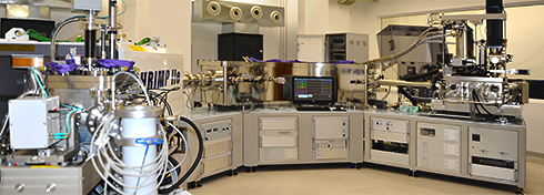
SHRIMP IIe/MC sensitive and high-resolution ion microprobe with multicollector SHRIMP instrument has been manufactured by Australian Scientific Instruments (ASI), with cooperation of the Australian National University in Canberra, where the ion microprobe was first designed and developed. SHRIMP is large radius, instrument capable of measuring the isotopic composition of low atomic number elements such as O, C, S using ceasium primary ion source and electron gun as well as high atomic number elements Pb, U, Th using duoplasmatron. The PGI ion microprobe is the fourth of the SHRIMP instruments in the world designed to a new enhanced configuration with two interchangeable primary sources: a duoplasmatron, for analyzing positive ions, and a Cs gun, for analyzing negative ions.
TECHNICAL PARAMETERS
The quality of the instrument can be assessed by its ability to detect trace elements present in the target at low concentrations (sensitivity) and its ability to distinguish between ions of very similar mass (mass resolution).
The high mass resolution of SHRIMP IIe is achieved by the use of a double-focusing mass spectrometer (energy and mass refocusing) with a very large turning radius of magnet and electrostatic analyzer (magnet radius 1000 mm, electrostatic analyzer radius 1272 mm).
Therefore SHRIMP has a beam line over 7 m long and weighs more than 13 tons.
The high sensitivity at high mass resolution: 5400 M/ΔM at 80% transmission with flat – topped peaks, permits resolution of major molecular interferences during analysis.
SHRIMP can be used for a routine measurement of the isotopic composition of the abundances of most elements in the Periodic Table and can operate in positive or negative ion mode.
SHRIMP is equipped with a Duoplasmatron for positive secondary ion analysis and a Caesium source and charge neutralization system for negative secondary ion analyses.
SHRIMP makes in situ isotopic and chemical analyses of complex solid materials (without chemical dissolution) by bombarding the sample surface with a high energy primary ion beam in a high vacuum, with a spot diameter of only a few microns.
This desorbs surface species through a physical process called sputtering. The primary ion beam diameter can be set from 30 microns to less than 5 microns.
The sputtered fragments are ionised, and then the secondary ions are gathered using electrostatic lenses and transferred to a mass spectrometer in which they are separated according to their relative masses.
The secondary mass analyser comprises an electrostatic sector, a quadrupole correcting lens and electromagnet. This design gives small values for all second-order image aberrations.
SAMPLE PREPARATION:
Mineral separation
Starting from crushing and grinding rocks to any fraction using heavy equipment, like jaw breakers and mills. Sieving the crushed rock through set of steel sieves of different diameter mesh. Washing out the dust from the sample in large beakers (sludge). Ending on extraction of selected minerals from the rocks: light, heavy, accessory, magnetic, non-magnetic and paramagnetic using Frantz isodynamic magnetic separator. Magnetic separation on Frantz is arguably the most essential method for bulk heavy mineral concentration; induced magnetic fields at various electric current settings.
Extraction such minerals as zircon, garnet, apatite, monazite, pyrochlore, rutile, and others minerals witch are not directly related to isotopic dating. Manual selection of minerals under binocular (stereomicroscope).
Mount preparation
Mount preparation should be done at Ion Microprobe Section to ensure specimen mounts are optimized for the SHRIMP. Room designed and equipped with world leading equipment for making mounts for ion microprobe investigations from typical samples such as mineral grains and microfossils to bigger ones in eg. human teeths
The majority of samples are embedded in epoxy disks. The end product is a 2.5 cm or 3.5 cm diameter circular mount that has been polished and includes mineral standards and labels. The equipment for mount preparation consists of precision cutter (Metkon Instruments Ltd.), semi-automatic grinding-polishing machine for preparing a wide variety of materials (Buehler, an ITW Company) and evaporative vacuum sputter Quorum Technologies Ltd for coating non-conductive samples with gold for ion microprobe analyses.
SAMPLE DOCUMENTATION BEFORE ANALYSIS
Careful imaging of polished mount is essential for analysis to see cracks, scratches, and some grain boundaries. Optical imaging is accomplished by Nikon Instrumentsmicroscope set designed for automatic and accurate imaging of every mount surface in transmitted and reflected light mode.
The sample documentation is also provided by new HITACHI SU 3500 scanning microscope equipped with Horiba Jobin Ivon -CL system.cathodoluminescence
SEM-CL allows effective image analyses with multiple signals:
1) a secondary electron SE providing surface rim information,
2) a back scattered electron BSE signal for compositional information and
3) a cathodoluminescence signal, which provide high resolution spectral information of luminescent materials and can be related to an intrinsic structural feature in the material or a specific elemental or/and isotopic composition.
The distribution of the CL signal in a material gives fundamental insights into such processes as crystal growth, replacement, dissolution features in igneous and metamorphic minerals deformation, provenance and some details of internal structures of fossils.
The new vacuum system of HITACHI SU 3500 enables various vacuum settings with range of 6-650 Pa. Low vacuum environment allows an observations of specimens in natural state and eliminates traditional sample preparation such as dehydratation and metal or carbon coating of non-conductive target. HITACHI SU3500 serves a clear, contrast-rich images starting from just 1.5 keV beam energy onwards. Thelow kV imaging can reveal fine surface details.
HITACHI SU3500 electron optics offers new class in imaging performance: an optional function for beam tilt based live display and acquisition of stereo image pairs even under FAST scanning, which can be observed with proper 3D glasses on the standard graphical user interface or in addition at an optional external 3D monitor.!


 PGI-NRI offer
 Mineral resources of Poland
  Oil and Gas in Poland


 Subscribe to RSS Feed